
Spinal tuberculosis, pathophysiology and radiological presentation, three case reports
Abstract
Prompt diagnosis and treatment of spinal tuberculosis are key in preventing its neurological and physical sequelae. This affection, also known as Pott’s disease, should be considered a differential diagnosis in patients presenting with unexplained back pain that can lead to neurological symptoms and eventually paraplegia.
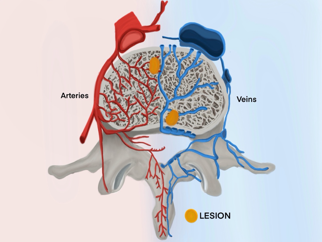
Mycobacterium tuberculosis, the etiological agent of tuberculosis, spreads from the lungs to the spine via venous or arterial pathways, causing lesions apparent upon imaging. Radiological findings include osseous destruction, disk collapse, abscess formation, and spinal deformity. While magnetic resonance is considered the most sensitive and specific imaging modality to establish a diagnosis, plain radiographs and computed tomography can provide useful information.
This manuscript discusses three Colombian cases of spinal tuberculosis with the goal of increasing familiarity regarding the pathophysiology, clinical and radiological manifestations, and differential diagnosis of this rare but potentially devastating disease.
Downloads
References
World Health Organization. Global tuberculosis report 2024. Geneva: World Health Organization; 2024. Accessed: August 26, 2024. Available at: https://iris.who.int/bitstream/handle/10665/379339/9789240101531-eng.pdf?sequence=1
Center for Disease Control and Prevention. Clinical overview of tuberculosis disease. Accessed: August 26, 2024. Available at: https://www.cdc.gov/tb/hcp/clinical-overview/tuberculosis-disease.html
Sharma SK, Mohan A, Kohli M. Extrapulmonary tuberculosis. Expert Rev Respir Med. 2021;15:931-48. https://doi.org/10.1080/17476348.2021.1927718
Garg RK, Somvanshi DS. Spinal tuberculosis: A review. J Spinal Cord Med. 2011;34:440-54. https://doi.org/10.1179/2045772311Y.0000000023
Stout J. Bone and joint tuberculosis. UpToDate. 2022. Accessed: August 26, 2024. Available at: https://www.uptodate.com/contents/search
World Health Organization. Global lists of high burden countries for tuberculosis (TB), TB/HIV and multidrug/rifampicin-resistant TB (MDR/RR-TB), 2021-2025: Background document. Geneva: World Health Organization; 2021. Accessed: August 26, 2024. Available at: https://iris.who.int/bitstream/handle/10665/341980/9789240029439-eng.pdf
World Bank: Incidence of tuberculosis. Accessed: August 26, 2024. Available at: https://data.worldbank.org/indicator/SH.TBS.INCD
Sternbach G. Percivall Pott: Tuberculous spondylitis. J Emerg Med. 1996;14:79-83. https://doi.org/10.1016/0736-4679(95)02053-5
Glassman I, Nguyen KH, Giess J, Alcantara C, Booth M, Venketaraman V. Pathogenesis, diagnostic challenges, and risk factors of Pott’s disease. Clin Pract. 2023;13:155-65. https://doi.org/10.3390/clinpract13010014
Semionov A, Lebel K, Diouf A, Pressacco J. Tuberculosis: A head-to-toe radiological review. Open J Radiol. 2022;12:207-21. https://doi.org/10.4236/ojrad.2022.124021
Manika K, Kipourou M, Georga S, Faniadou E, Pilianidis G, Arsos G, et al. 18F-FDG PET/CT contribution to tuberculous vertebral osteomyelitis diagnosis: a case report. Oxf Med Case Rep. 2020;9. https://doi.org/10.1093/omcr/omaa068
Shah LM, Salzman KL. Imaging of spinal metastatic disease. Int J Surg Oncol. 2011;10. https://doi.org/10.1155/2011/769753
Tu L, Liu X, Gu W, Wang Z, Liu Z, Zhang E, et al. Imaging-assisted diagnosis and characteristics of suspected spinal brucellosis: A retrospective study of 72 cases. Med Sci Monit. 2018;24:2647-54. https://doi.org/10.12659/MSM.909288
Rizkalla JM, Alhreish K, Syed IY. Spinal brucellosis: A case report and review of the literature. J Orthop Case Rep. 2021;11:1-5 https://doi.org/10.13107/jocr.2021.v11.i03.2060
Some similar items:
- Juan Gabriel Bueno-Sánchez, Jairo René Martínez-Morales, Elena E. Stashenko, Wellman Ribón, Anti-tubercular activity of eleven aromatic and medicinal plants occurring in Colombia , Biomedica: Vol. 29 No. 1 (2009)
- María Consuelo Garzón, Dailyn Yorledy Angée, Claudia Llerena, Dora Leticia Orjuela, Jorge Ernesto Victoria, Surveillance of Mycobacterium tuberculosis resistance to antituberculosis drugs , Biomedica: Vol. 28 No. 3 (2008)
- Diego Chaves, Andrea Sandoval, Luis Rodríguez, Juan C. García, Silvia Restrepo, María Mercedes Zambrano, Comparative analysis of six Mycobacterium tuberculosis complex genomes , Biomedica: Vol. 30 No. 1 (2010)
- María Imaz, Sonia Allassia, Mónica Aranibar, Alba Gunia, Susana Poggi, Ana Togneri, Lidia Wolff, Group of Implementation of Fluorescence, Performance of LED fluorescence microscopy for the detection of acid-fast bacilli from respiratory samples in peripheral laboratories in Argentina , Biomedica: Vol. 37 No. 2 (2017)
- Diana Castaño, Mauricio Rojas, Alterations in recruitment and activation of Rab proteins during mycobacterial infection , Biomedica: Vol. 30 No. 2 (2010)
- Adriana Rojas-Villarraga, Carlos Andrés Agudelo, Ricardo Pineda-Tamayo, Alvaro Porras, Gustavo Matute, Juan Manuel Anaya, Tuberculosis in patientes treated with tumor necrosis factor alpha antagonists living in an endemic area. Is the risk worthwhile? , Biomedica: Vol. 27 No. 2 (2007)
- Carlos A. Torres-Duque, Claudia Díaz, Leslie Vargas, Elsa María Serpa, Walter Mosquera, María Consuelo Garzón, Graciela Mejía, Luz Mary García, Liliana Andrea González, Claudia Marcela Castro, Wellman Ribón, Disseminated mycobacteriosis affecting a prosthetic aortic valve: first case of Mycobacterium peregrinum type III reported , Biomedica: Vol. 30 No. 3 (2010)
- Francisco Cuervo, Luis F. Giraldo, Alirio Bastidas, Carlos Vélez, Maria R. Forero, Fibrinogen-thrombin as bridge therapy in massive hemoptysis , Biomedica: Vol. 33 No. 1 (2013)
- Alvaro Javier ldrovo, Notes on the onset of a pulmonary tuberculosis epidemic in Bogotá (1870-1920) , Biomedica: Vol. 21 No. 3 (2001)
- Ivohne Fernanda Corrales, Jorge Alberto Cortés, María Lucía Mesa, Graciela Zamora, Sternal osteomyelitis and scrofuloderma due to BCG vaccination. , Biomedica: Vol. 23 No. 2 (2003)
Copyright (c) 2025 Biomedica

This work is licensed under a Creative Commons Attribution 4.0 International License.
| Article metrics | |
|---|---|
| Abstract views | |
| Galley vies | |
| PDF Views | |
| HTML views | |
| Other views | |