

A novel activity on thymocytes cells exerted by the rattlesnake (Crotalus durissus cumanensis) venom
Abstract
Introduction: The thymus is active mainly during the neonatal and pre-adolescent periods.
Objective: To test naïve thymocytes proliferation and monocytes stimulation.
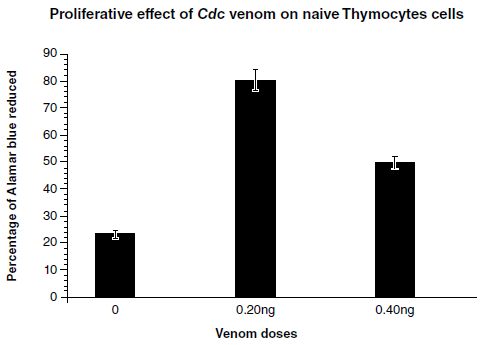
Materials and methods: We collected fresh thymus tissue from neonate mice after surgery. Suspension cells were coated onto Ficoll-Hypaque support. The obtained cells (thymocytes) were cultured measuring the proliferation of naïve T cells stimulated by Crotalus durissus cumanensis (Cdc) venom at sub-lethal doses (20 ng). Then, we supplemented the wells with AlamarBlue™ and incubated them for 5 h to test their proliferation. Mononuclear cells from mice peripheral blood were collected and layered onto the support of the Ficoll-Hypaque solution. We added the thymocytes actively dividing (25 x 105 cells) from cultures stimulated with Cdc venom at 20 ng/well to cultured monocytes freshly obtained from the Ficoll-Hypaque separation. Both cell populations were incubated for 36 h until monocytes matured to macrophages.
Results: The naïve thymocytes rapidly proliferated after stimulation with the Cdc venom (NTCdc) and these successively induced the maturation and function of monocytes progenitor cells to mature macrophages, which ingested Chinese ink.
Conclusions: The naïve thymocytes proliferated by stimulation with the Cdc venom and subsequently the NT/Cdc induced the rapid maturation and function of monocytes progenitor cells becoming mature macrophages with their phenotypic characteristics.
Downloads
References
Markert ML, Devlin BH, Chinn IK, McCarthy EA. Thymus transplantation in complete DiGeorge anomaly. Immunol Res. 2009;44:61-70. https://doi.org/10.1007/s12026-008-8082-5
Hernández-Cruz S, García-Jiménez R, Zucatelli-Mendonc A, Petricevich VL. Pro- and antiinflammatory cytokines release in mice injected with Crotalus durissus terrificus venom. Mediators Inflamm. 2008;2008:874962. https://doi.org/10.1155/2008/874962
Holland AM, van den Brink MR. Rejuvenation of the aging T cell compartment. Curr Opin Immunol. 2009;21:454-9. https://doi.org/10.1016/j.coi.2009.06.002
van den Broek T, Borghans JAM, van Wijk F. The full spectrum of human naive T cells. Nat Rev Immunol. 2018;18:363-73. https://doi.org/10.1038/s41577-018-0001-y
van den Brink MR, Velardi E, Perales MA. Immune reconstitution following stem cell transplantation. Hematology Am Soc Hematol Educ Program. 2015;2015:215-9.
https://doi.org/10.1182/asheducation-2015.1.215
Tominaga K, Kinoshita Y, Hato F, Masudå A, Matsuyama M. Effects of cholinergic agonists on the proliferation and protein synthesis in a cultured thymic epithelial cell line. Cell Mol Biol. 1989;35:679-86.
Chang C. Looking back on the discovery of alpha-bungarotoxin. J Biomed Sci. 1999;6:368-75. https://doi.org/10.1007/BF02253668
National Research Council (US) Institute for Laboratory Animal Research. Guide for the Care and Use of Laboratory Animals. Washington, D.C.: National Academies Press; 1996.
Stadnyk AW, Befus AD, Gauldie J. Characterization of nonspecific esterase activity in macrophages and intestinal epithelium of the rat. J Histochem Cytochem. 1990;38:1-6. https://doi.org/10.1177/38.1.1688447
Nakul-Aquaronne D, Bayle J, Frelin C. Coexpression of endothelial markers and CD14 by cytokine mobilized CD34+ cells under angiogenic stimulation. Cardiovasc Res. 2003;57:816-23. https://doi.org/10.1016/s0008-6363(02)00776-9
Gowans JL, Gesner BM, McGregor DD. The immunological activity of lymphocytes. In: Wolstenholme GEW, O’Connor M, editors. Biological activity of the leucocyte. London, England: Ciba Foundation Study Group; 1961. p. 32.
Miller JFAP: Effect of neonatal thymectomy on the immunological responsiveness of the mouse. Proc Roy Soc London. 1962;156B:410-28.
Miller JFAP. Immunological function of the thymus. Lancet. 1961;2:748-9. https://doi.org/10.1016/s0140-6736(61)90693-6
Miller JFAP: Aetiology and pathogenesis of mouse leukaemia. Adv Cancer Res 1961;6:291-368. https://doi.org/10.1016/s0065-230x(08)60623-5
Beard J. The source of leucocytes and the true function of the thymus. Anat Anz. 1990;18:550-60.
Gordon S, Taylor PR. Monocyte and macrophage heterogeneity. Nat Rev Immunol. 2005;5:953-64. https://doi.org/10.1038/nri1733
Murray PJ, Wynn TA. Protective and pathogenic functions of macrophage subsets. Nat Rev Immunol. 2011;11:723-37. https://doi.org/10.1038/nri3073
Muftuoglu TM, Koksal N, Ozkutlu D. Evaluation of phagocytic function of macrophages in rats after partial splenectomy. J Am Coll Surg. 2000;191:668-71. https://doi.org/10.1016/s1072-7515(00)00739-0
Bredenkamp N, Nowell CS, Blackburn CC. Regeneration of the aged thymus by a single transcription factor. Development. 2014;141:1627. https://doi.org/10.1242/dev.103614
Rosenblum MD, Gratz IK, Paw JS, Abbas AK. Treating human autoimmunity: current practice and future prospects. Sci Transl Med. 2012;4:125sr1. https://doi.org/10.1126/scitranslmed.3003504
Hur J, Yoon CH, Kim HS, Choi JH, Kang HJ, Hwang KK, et al. Characterization of two types of endothelial progenitor cells and their different contributions to neovasculogenesis. Arterioscler Thromb Vasc Biol. 2004;24:288-93. https://doi.org/10.1161/01.ATV.0000114236.77009.06
Vergadi E, Ieronymaki E, Lyroni K, Vaporidi K, Tsatsanis C. Akt signaling pathway in macrophage activation and M1/M2 polarization. J Immunol. 2017;198:1006-14. https://doi.org/10.4049/jimmunol.1601515
Some similar items:
- Jacqueline Barona, Rafael Otero, Vitelbina Programa de Ofidismo/Escorpionismo, Facu Núñez, Toxicological and immunological aspects of scorpion venom (Tytius pachyurus): neutralizing capacity of antivenoms produced in Latin America. , Biomedica: Vol. 24 No. 1 (2004)
- Vera María Ripoll, David Hume, Marta Raquel Fontanilla, Specific expression of inflammatory genes in macrophage subpopulations. , Biomedica: Vol. 25 No. 2 (2005)
| Article metrics | |
|---|---|
| Abstract views | |
| Galley vies | |
| PDF Views | |
| HTML views | |
| Other views | |
Funding data
-
Consejo de Desarrollo Científico y Humanístico, Universidad Central de Venezuela
Grant numbers PG: 09-8760-2013