

Pheohyphomycosis skin nodule in a young woman
Abstract
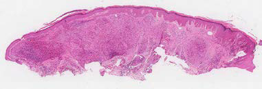
We present a 28 year-old woman with a five-year history of an asymptomatic slowly growing 10 mm nodule on her right thigh, with clinical features suggestive of either a dermatofibroma or a keloid.
The nodule was excised, and histopathological examination revealed prominent dermal granulomas containing numerous giant cells, focal microabscesses, abundant pigmented yeasts with dark walls –some arranged in chains–, and septate hyphae with blackish walls, findings initially suggestive of chromoblastomycosis. The abundance of moniliform hyphae arranged in linear chains allowed us to diagnose cutaneous pheohyphomycosis without hypodermal invasion.
We highlight the chronic nature of the condition, its localized presentation, and the prominence of granulomas rich in giant cells –with scarce abscesses– as notable findings.
The patient’s clinical course remains unknown, as she did not return for follow-up.
Downloads
References
1. Rinaldi MG. Phaeohyphomycosis. Dermatol Clin 1996;14:147-53. https://doi.org/10.1016/s0733-8635(05)70335-1
2. De Hoog GS, Queiros-Tellez F, Haase G, Fernandez-Zeppenfeldt G, Angelis AT, et al. Black fungi: Clinical and pathogenic approaches. Med Mycol. 2000;38(Suppl.1):243-50.
3. Ajello L. Phaeohyphomycosis: Definition and etiology, Mycosis. Scientific publication No. 304. Washington, D.C.: Pan American Health Organization; 1986. p. 16-130.
4. Peltroche-Llacsahuanga H, Schnitzler N, Jentsch S, Platz A, De Hoog GS, Schweitzer KG, et al. Analyses of phagocytosis, evoked oxidative burst, and killing of black yeasts by human neutrophils: A tool for estimating their pathogenicity? Med Mycol 2003;41:7-14. https://doi.org/10.1080/mmy.41.1.7.14
5. Revankar SG. Phaeohyphomycosis. Infect Dis Clin North Am. 2006;20:609-20. https://doi.org/10.1016/j.idc.2006.06.004
6. Vitale RG, De Hoog GS. Molecular diversity, new species, and antifungal susceptibilities in the Exophiala spinifera clade. Med Mycol. 2002;40:545-56. https://doi.org/10.1080/mmy.40.6.545.556
7. Elias Costa MR, Da Silva Lacaz C, Kawasaki M, De Camargos ZP. Conventional versus molecular diagnostic tests. Med Mycol. 2000;38(Suppl.1):139-45.
8. Queiros-Tellez F, Nucci M, Lopes CA, Tobón A, Restrepo A. Mycoses of implantation in Latin America: An overview of epidemiology, clinical manifestations, diagnosis, and treatment. Med Mycol. 2011;49:225-36. https://doi.org/10.3109/13693786.2010.539631
9. Rippon JW. Faeohifomicosis. En: Tratado de Micología Médica. 3a edición. México, D. F.: McGraw Hill; 1990. p 321-49.
10. Bonifaz A. Micología Médica Básica. 2ª edición. México, D.F.: Méndez Editors; 2003.
11. Rodríguez-Toro G, Palencia Y. Esporotricosis. Valor diagnóstico del cuerpo asteroide. Biomédica. 1985;5:41-6. https://doi.org/10.7705/biomedica.v5i1-2.1900
12. Frasquet-Artés JS, Pemán J, Blanes M, Hernández-Porto M, Cano J, et al. Feohifomicosis cerebral: descripción de un caso y revisión de la literatura. Rev Iberoam Micol. 2014;31:197-202. https://doi.org/10.1016/j.riam.2012.12.008
13. Alayeto Ortega J, Alier Fabregó A, Puig Verdie L, Sorli Redo ML, Horcajada Gallego JP, Portillo Bordonabe ME. Feohifomicosis subcutánea causada por Phaeoacremonium parasiticum. Rev Iberoamer Micol. 2015;32:265-8. https://doi.org/10.1016/j.riam.2014.10.004
14. Scupsky H, Junkins-Hopkins J. Counterfeit pennies: Distinguishing chromoblastomycosis from phaeohyphomycotic infections. Am J. Dermatopatol. 2017;39:485-7. https://doi.org/10.1097/DAD.0000000000000679
15. Fariñas MC, Fernández-Sampedro M, Armiñanzas C. Formas clínicas y tratamiento de las infecciones causadas por otros hongos filamentosos. Enferm Infecc Microbiol Clin. 2012;30:414-9.
16. Feng P, Najafzadeh M, Sun J, Ahmed S, Xi L, de Hoog GS, et al. In vitro activities of nine antifungal drugs against 81 Phialophora and Cyphellophora isolates. Antimicrob Agents Chemother. 2012;56:6044-7. https://doi.org/10.1128/AAC.01112-12
Some similar items:
- Guillermo Sánchez, John Nova, Reliability and reproducibility of the Fitzpatrick phototype scale for skin sensitivity to ultraviolet light , Biomedica: Vol. 28 No. 4 (2008)
- Angélica Ballesteros, Sandra Beltrán, Jaime Patiño, Cynthia Bernal, Rocío Orduz, Disseminated juvenile paracoccidioidomycosis diagnosed in a girl in an urban area , Biomedica: Vol. 34 No. 1 (2014)
- John Fredy Nieto-Ríos, Douglas Ramón Villafañe-Bermúdez, Gustavo Adolfo Guerrero-Tinoco, Isabel Cristina Ramírez-Sánchez, Lina María Serna-Higuita, Arbey Aristizábal-Alzate, Catalina Ocampo-Kohn, Gabriel Varela, Gustavo Zuluaga-Valencia, Brain abscess caused by Cladophialophora bantiana after renal allograft loss: A case report , Biomedica: Vol. 39 No. Supl. 2 (2019): Enfermedades transmisibles en el trópico, agosto
- Paola Macías , Juliana Ordóñez , Claudia M. Arenas , Gerzaín Rodríguez , An 18-year-old man with tropical verrucous syndrome: Leishmaniasis or sporotrichosis? , Biomedica: Vol. 41 No. 2 (2021)
- Ana María Sanín , Ángela María Londoño , Verónica Gil , Ana María Mejía , Hernán Darío Aguirre, Elsa María Vásquez , Catalina Valencia , Carolina Cardona, Mucocutaneous manifestations and their relationship with CD4 T-lymphocyte count in hospitalized patients infected with the human immunodeficiency virus (HIV) in Medellín, Colombia , Biomedica: Vol. 42 No. 2 (2022)
- Mauricio Torres, Juliana Flórez, María Salomé Páez, Ángela María Londoño, Paola Cárdenas , Mariela Tavera, Mónica Paola Novoa , Carolina Cortés, Rosángela Casanova, Pediatric psoriasis: A descriptive, retrospective and multicenter study in Colombia , Biomedica: Vol. 45 No. 2 (2025)
- Manuel Calvopina, Elías Guamán-Charco , Jasmín Vélez , Belén Vélez , Camila González , Hemorraghic skin syndrome associated with contact of Lonomia spp. caterpillar: First report from the Ecuadorian Amazon , Biomedica: Vol. 45 No. 3 (2025)
Copyright (c) 2025 Biomedica

This work is licensed under a Creative Commons Attribution 4.0 International License.
| Article metrics | |
|---|---|
| Abstract views | |
| Galley vies | |
| PDF Views | |
| HTML views | |
| Other views | |