

Septo-optic dysplasia plus: A case report for reviewing and recognizing this condition
Abstract
Septo-optic dysplasia is a congenital neurological condition with multifactorial etiology, characterized by septum pellucidum agenesis and/or corpus callosum dysgenesis, hypoplasia of the chiasm or optic nerves, and hormonal dysfunction with pituitary or hypothalamic alterations. Diagnosis requires two of these criteria and magnetic resonance is the imaging test of choice. Most cases present with abnormalities of cortical development in the form known as septo-optic dysplasia plus. While seizures and neurodevelopmental disorders are the dominant neurological manifestations, this entity is highly heterogeneous and has multiple clinical and radiological findings to consider.
We present the case of a 35-year-old man with a history of cranioencephalic trauma in childhood and remission for refractory focal epilepsy associated with cognitive deficit.
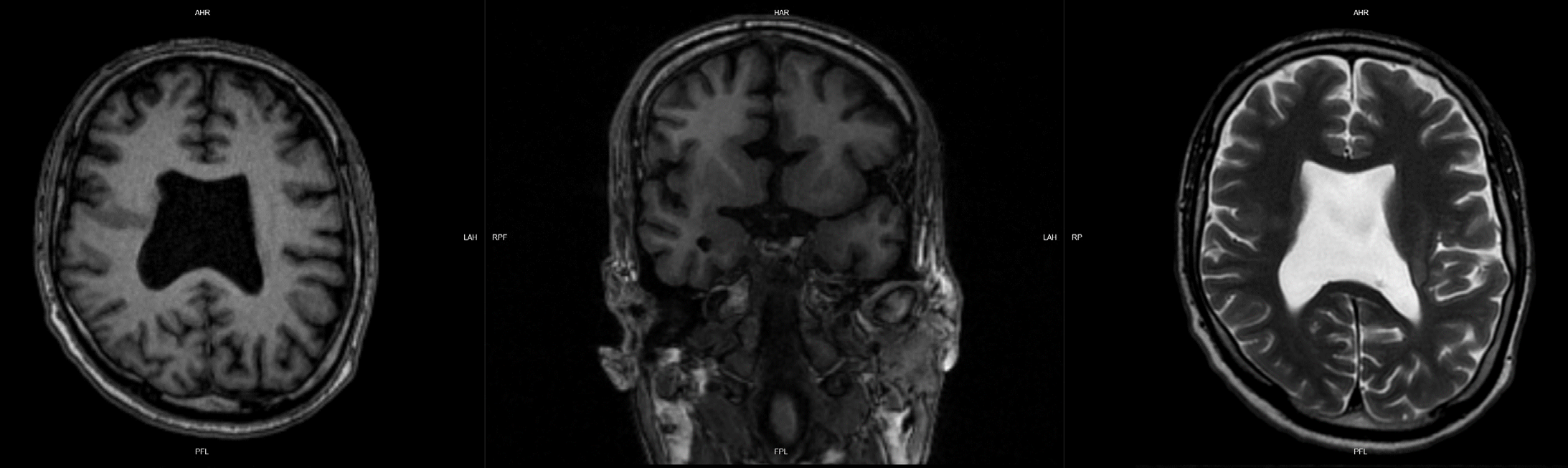
During the initial examination, the simple cranial tomography showed septum pellucidum agenesis and corpus callosum dysgenesis. Brain magnetic resonance imaging revealed agenesis of the septum pellucidum, irregularity and anomalous thickening of the cerebral cortex in frontal lobes and perisylvian region, heterotopic gray matter in frontal lobes and left fronto-insular region, mild supratentorial ventriculomegaly, atypical appearance of the corpus callosum rostrum, and hypoplasia of the chiasm and optic nerves.
Although agenesis of the septum pellucidum was the key finding in this case, it is not present in all patients. The relevance of magnetic resonance imaging for the detailed evaluation of other involved structures, highlighting optic nerve hypoplasia, is fundamental in the radiologist’s diagnostic workup and this entity recognition.
Downloads
References
Ferran Kd, Paiva IA, Gilban DL, Resende M, Souza MA, Beserra IC, et al. Septo-optic dysplasia. Arq Neuropsiquiatr. 2010;68:400-5. https://doi.org/10.1590/s0004-282x2010000300014
Cerbone M, Güemes M, Wade A, Improda N, Dattani M. Endocrine morbidity in midline brain defects: Differences between septo-optic dysplasia and related disorders. eClinMed. 2020;19:100224. https://doi.org/10.1016/j.eclinm.2019.11.017
Zoric L, Nikolic S, Stojcic M, Zoric D, Jakovljevic S. Septo-optic dysplasia plus: A case report. BMC Res Notes. 2014;7:191. https://doi.org/10.1186/1756-0500-7-191
Severino M, Allegri AE, Pistorio A, Roviglione B, Di Iorgi N, Maghnie M, et al. Midbrain-hindbrain involvement in septo-optic dysplasia. AJNR Am J Neuroradiol. 2014;35:1586-92. https://doi.org/10.3174/ajnr.a3959
Khokhar A, Umpaichitra V, Pérez-Colón S. Septo-optic dysplasia among children in Central Brooklyn. Ann Pediatr Child Health. 2015;3:1076. https://doi.org/10.47739/2373-9312/1076
Barkovich AJ, Fram EK, Norman D. Septo-optic dysplasia: MR imaging. Radiology. 1989;171:189-92. https://doi.org/10.1148/radiology.171.1.2928524
Gasparetto EL, Warszawiak D, de Carvalho Neto Ad, Benites Filho PR, Bruck I, Antoniuk S. Septo-optic dysplasia plus: Case report. Arq Neuropsiquiatr. 2003;61:671-6. https://doi.org/10.1590/s0004-282x2003000400028
Mncube SS, Goodier MD. Normal measurements of the optic nerve, optic nerve sheath and optic chiasm in the adult population. SA J Radiol. 2019;23:1772. https://doi.org/10.4102%2Fsajr.v23i1.1772
Wagner AL, Murtagh FR, Hazlett KS, Arrington JA. Measurement of the normal optic chiasm on coronal MR images. AJNR Am J Neuroradiol. 1997;18:723-6. Erratum in: AJNR Am J Neuroradiol 1997;18:1396.
Al-Senawi R, Al-Jabri B, Al-Zuhaibi S, Al-Azri F, Al-Yarubi S, Harikrishna B, et al. Septooptic dysplasia complex: Clinical and radiological manifestations in Omani children. Oman J Ophthalmol. 2013;6:193-8. https://doi.org/10.4103%2F0974-620X.122277
Ward DJ, Connolly DJA, Griffiths PD. Review of the MRI brain findings of septo-optic dysplasia. Clin Radiol. 2021;76:160.e1-160.e14. https://doi.org/10.1016/j.crad.2020.09.007
Miller SP, Shevell MI, Patenaude Y, Poulin C, O’Gorman AM. Septo-optic dysplasia plus: A spectrum of malformations of cortical development. Neurology. 2000;54:1701-3. https://doi.org/10.1212/wnl.54.8.1701
Kuban KC, Teele RL, Wallman J. Septo-optic-dysplasia-schizencephaly. Radiographic and clinical features. Pediatr Radiol. 1989;19:145-50. https://doi.org/10.1007/bf02388642
Gutiérrez-Castillo A, Jiménez-Ruiz A, Chávez-Castillo M, Ruiz-Sandoval JL. Septo-optic dysplasia plus syndrome. Cureus. 2018;10:e3727. https://doi.org/10.7759/cureus.3727
Sener RN. Septo-optic dysplasia associated with cerebral cortical dysplasia (cortico-septooptic dysplasia). J Neuroradiol. 1996;23:245-7.
AlKhateeb M, McLachlan R, Burneo J, Diosy D, Mirsattari S. Six adult patients with septooptic dysplasia and drug-resistant epilepsy: Clinical findings and course. Epilepsy Behav Case Rep. 2017;8:73-84. https://doi.org/10.1016/j.ebcr.2017.04.001
Alt C, Shevell MI, Poulin C, Rosenblatt B, Saint-Martin C, Srour M. Clinical and radiologic spectrum of septo-optic dysplasia: Review of 17 cases. J Child Neurol. 2017;32:797-803. https://doi.org/10.1177/0883073817707300
Leventer RJ, Guerrini R, Dobyns WB. Malformations of cortical development and epilepsy. Dialogues Clin Neurosci. 2008;10:47-62. https://doi.org/10.31887/dcns.2008.10.1/rjleventer
Desikan RS, Barkovich AJ. Malformations of cortical development. Ann Neurol. 2016;80:797-810. https://doi.org/10.1002/ana.24793
Kuriyama M, Shigematsu Y, Konishi K, Konishi Y, Sudo M, Haruki S, et al. Septo-optic dysplasia with infantile spasms. Pediatr Neurol. 1988;4:62-5. https://doi.org/10.1016/0887-8994(88)90028-8
Carrascosa-Romero MC, Ruiz-Cano R, Martínez-López F, Alfaro-Ponce B, Pérez-Pardo A. Midriasis congénita como signo inicial de displasia septo-óptica. Arch Soc Esp Oftalmol. 2013;88:398-402. https://doi.org/10.1016/j.oftal.2012.05.005
Levine LM, Bhatti MT, Mancuso AA. Septo-optic dysplasia with olfactory tract and bulb hypoplasia. J AAPOS. 200;5:398-9. https://doi.org/10.1067/mpa.2001.118869
Gündüz K, Günalp I, Saatçi I. Septo-optic dysplasia associated with bilateral complex microphthalmos. Ophthalmic Genet. 1996;17:109-13. https://doi.org/10.3109/13816819609057113
Dhingra M. Uncommon presentation of septo-optic dysplasia: Case report. Endocrinol Metab Int J. 2017;4:129-31. https://doi.org/10.15406/emij.2017.04.00102
Lobo AR, Ocampo M. Septo-optic dysplasia a case presentation for revisiting this intriguing and uncommon condition. J Radiol Med Imaging. 2021:4;1044.
Some similar items:
- Sara Emilia Giraldo, Javier Rincón, Pilar Puebla, Mariel Marder, Cristina Wasowski, Nadezdha Vergel, Mario Francisco Guerrero, Isovaleramide, an anticonvulsant molecule isolated from Valeriana pavonii , Biomedica: Vol. 30 No. 2 (2010)
- Blair Ortiz, Yesyka Jaramillo, Christian Rojas, X-linked epileptic syndrome by protocadherin 19 mutation associated with leukoencephalopathy and posterior reversible tractopathy , Biomedica: Vol. 38 No. 4 (2018)
- David Ríos, Carlos Cárdenas, Patricia Quintero, Dyke-Davidoff-Masson syndrome: Adult female patient with refractory epilepsy and global cognitive decline , Biomedica: Vol. 45 No. 2 (2025)
Copyright (c) 2024 Biomedica

This work is licensed under a Creative Commons Attribution 4.0 International License.
| Article metrics | |
|---|---|
| Abstract views | |
| Galley vies | |
| PDF Views | |
| HTML views | |
| Other views | |